OBJECTIVE:
By the end of the exercise you should be able to:
1. Describe the events associated with the cell cycle.
2. Describe the events associated with mitosis.
3. Distinguish the phases of mitosis on prepared slides of mitotic cells.
4. Outline the events of each stage of meiosis.
5. Compare and contrast meiosis with mitosis.
6. Discuss the relevance of meiosis to sexual reproduction.
7. Discuss the relationship of meiosis and gametogenesis.
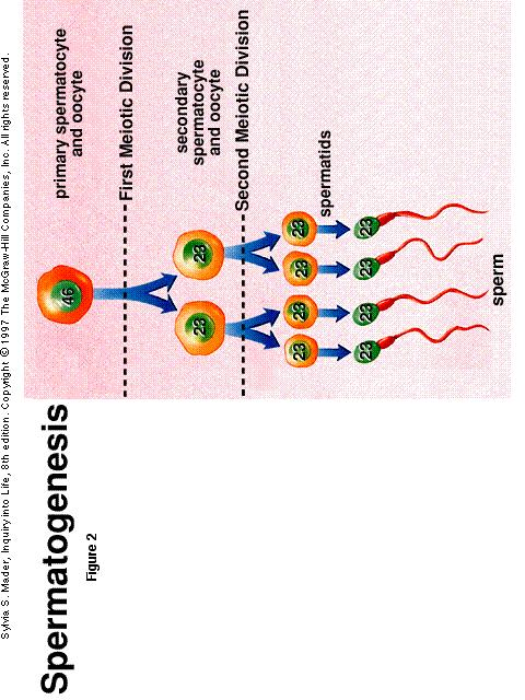
8. Describe the events of spermatogenesis and oogenesis.
INTRODUCTION:
The remarkable diversity of form and function that eukaryotic cells assume is even more remarkable when you consider that a multicellular organism begins life as a single cell, the zygote. The egg and sperm (the gametes), though usually unequal in size, give an equal number of chromosomes to the zygote: each contributes the haploid number of chromosomes. The developing organism, therefore, contains a diploid number of chromosomes - a haploid set from each parent. For example, the diploid complement of a human zygote (and subsequently all other body cells) is 46 chromosomes; the haploid complement of each gamete is 23 chromosomes. After fertilization, the zygote gives rise to all the cells that make up the organism by repeated cell divisions, called mitosis. In multicellular organisms, mitosis permits growth and repair of tissues. In eukaryotic unicellular organisms, mitosis is a form of asexual reproduction.
During the formation of the eggs and sperm, the
chromosome number is reduced from diploid to haploid by a type of cell
division called meiosis. We will examine this form of cell division later
in this laboratory exercise.
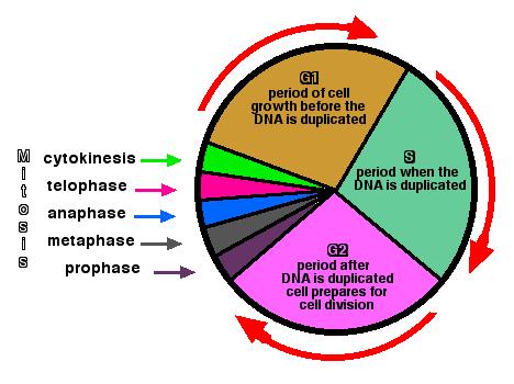
A. THE CELL CYCLE:
The complex series of events that encompasses the life span of an actively dividing cell is termed the cell cycle, which includes an interphase during which copy of the DNA) occurs, as well as the synthesis of RNA and proteins. Note that replication of DNA occurs only during a period of interphase called the S phase (S for "synthesis"). The doubling of DNA during the S phase provides a full complement of DNA for the 2 daughter cells that will result from the ensuing mitotic division. During interphase, there are also 2 phases called G1 and G2. (G for "gap"). The G1 phase, which precedes DNA replication, includes a variety of growth processes and synthesis of compounds other than DNA. During the G2 phase, which immediately follows DNA replication, the molecules. and structures directly involved with mitosis are synthesized, assembled. The combination of G1, S, G2, and M phases make up the cell cycle (mitotic cycle).
Mitosis is essentially the same in all organisms
but, just as plant and animal cells differ to some extent structurally,
there are some differences between them in this process. It is the objective
of this part of the lab exercise to examine the essential steps in mitosis
and to characterize the similarities and differences in this process between
plant and animal cells.
1. MITOSIS IN PLANTS CELLS
The onion (Allium) root tip is one of the most widely used materials for the study of mitosis because it is readily available, preparation of the dividing cells is easy, and the chromosomes are large and few in number. Root tips of plants contain meristems, which are localized areas of rapid cell division; therefore, chances are good that in a specimen of such tissues, one can find every stage of mitosis.
Obtain a slide of onion root tips, and note a series of dark streaks on it. Each streak is a very thin longitudinal section through an onion root tip.
Place the slide on the stage of your compound microscope, and locate 1 of the sections under low power ( I0X). It is often possible to determine whether a given section shows good examples of mitotic stages. Because each section is very thin, not all will be equally good for study. After your preliminary examination under low power, change to high power (40X or 43X), being careful not to break the slide. Keep in mind the sequence in which the stages occur, but do not try to find them in sequence. Thus, if you happen to find an anaphase first, study it before proceeding to another stage. Because cells remain in interphase and prophase longer than in the other stages, chances are that most of the cells will be in interphase; many will be in prophase; and only a few will be in the other stages.
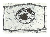
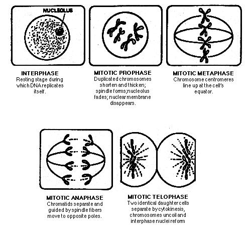
a. Interphase
The interphase cell, so named because early biologists
thought it was a resting phase, is actively undergoing cellular respiration
and the synthesis of DNA, RNA, and protein. Interphase cells are characterized
by a distinct nucleus bounded by a nuclear envelope. One or more nucleoli
may be visible.
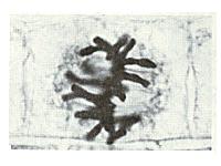
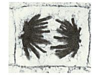
d. Anaphase
Anaphase begins when the sister chromatids are
pulled apart by the spindle fibers; the centromere splits, and the sister
chromatids become daughter chromosomes as they are pulled toward opposite
poles of the cell. This stage can be recognized in the onion cell by the
2 groups of V-shaped chromosomes; the sharp end of the V is oriented toward
the pole. The onion has a diploid number of 16 chromosomes, so it is seldom
possible to see all of them at 1 time.
Reduce the light by adjusting the diaphragm of your microscope, and see if you can find any spindle fibers near the center of the cell. They appear as very fine lines between the 2 groups of chromosomes, but they are often not visible in a study of this kind. Anaphase ends when the newly-separated daughter chromosomes arrive at opposite poles of the cell.
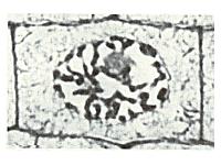
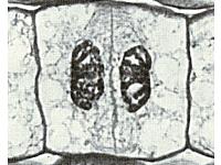
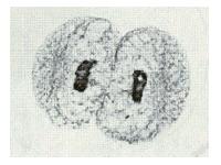
e. Telophase
.It is often difficult to distinguish late anaphase
from early telophase in the cells of the onion root tip. As telophase
progresses, the nuclei begin to reorganize, the chromosomes uncoil and
become longer and thinner, the nuclear envelope reforms by fusion of parts
of the endoplasmic reticulum, and the nucleoli reappear. Mitosis ends with
the assembly of 2 interphase nuclei, each with 1 complete set of single-stranded
chromosomes.
f. Cytokinesis
Mitosis is completed during cytokinesis.
The first indication that cytokinesis is beginning, a cell plate.starts
to form as a fine line across the center of the cell. When complete the
cell plate divides the original cell into 2 daughter cells. In some cells,
the cell plate is indistinct. The daughter cells resulting from mitotic
division have the same number and kinds of chromosomes as the original
cell. Thus, in the onion, each daughter cell has 16 chromosomes.
2. MITOSIS IN ANIMAL CELLS
Mitosis in animal cells is easily observed on a prepared slide of a whitefish blastula. (A blastula is an early stage of development of an embryo formed by successive mitotic divisions.)
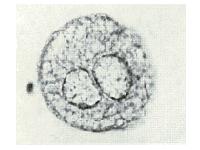
a. Interphase
As in plant cells, animal cells in interphase are
characterized by a distinct nucleus bounded by a nuclear envelope. The
nucleolus should be identifiable. Immediately adjacent to the nuclear envelope
is a cytoplasmic organelle known as the centrosome.
b. Prophase
As in plant cells, mitosis begins with prophase.
During prophase, in contrast to plant cells, 2 pairs of structures called
centrioles form within the centrosome. They begin to move apart, migrating
around the nucleus toward the opposite poles of the cell. Microtubules
radiate from each pair of centrioles like spokes on a wheel, forming a
configuration known as an aster. The centrioles continue to migrate until
they lie at opposite poles of the cell. When the nuclear envelope disintegrates,
the spindle becomes visible between the centrioles. Locate various stages
of prophase on the slide.
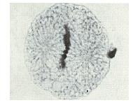
c. Metaphase
During metaphase, the chromosomes move toward the
central region of the spindle to form the metaphase plate. By the time
the chromosomes are organized along the metaphase plate, the nuclear envelope
has completely disintegrated. Locate this phase of mitosis on your slide.
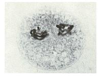
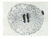
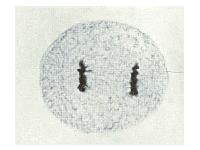
d. Anaphase
The sister chromatids that make up each chromosome
are now separated from each other and are pulled by the spindle fibers
to opposite poles of the cell. The separated sister chromatids now become
daughter chromosomes. When the daughter chromosomes reach the poles, anaphase
ends and telophase begins.
Meiosis occurs in all sexually reproducing eukaryotes; it is the type of cell division that is involved in the formation of eggs and sperm (gametes). The formation of gametes is called gametogenesis. In the process of meiosis, the diploid number of chromosomes is reduced to the haploid number; thus meiosis is sometimes called "reduction division". In evolutionarily advanced organisms, chromosomes are diploid (2N); i.e., they occur as pairs in each nucleus. The 2 chromosomes of a pair are called homologous chromosomes, or homologous; each homologue of a pair has the same sites or loci for the same genes. Homologous chromosomes are not identical they contain variants of the same genes.
The reduction from the diploid number to the haploid number of chromosomes that takes place in meiosis is significant, because a cell with a haploid number of chromosomes (i.e., a gamete) can fuse with another haploid cell (i.e., another gamete) during sexual reproduction and restore the original diploid number of chromosomes to the resulting zygote. Thus, meiosis keeps the chromosome number constant for most species of plants and animals. In addition to reducing the number of chromosomes, meiosis shuffles the genetic material so that each resulting cell carries a new and unique set of genes.
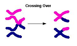
As in mitosis, meiosis is preceded by each chromosome replicating to form 2 chromatids attached at a centromere. However, there are 2 important events that occur in meiosis that do not occur in mitosis: 1) Meiosis consists of 2 rounds of chromosome separation into daughter cells; chromosomes are replicated in the first round but not in the second. Thus, the genetic material is replicated once and divided twice; this produces 1/2 the original number of chromosomes. 2) In an early stage of meiosis, each chromosome (composed of 2 sister chromatids) pairs along its length with its homologue. This pairing of homologous is called synapsis; during which the 4 chromatids (known collectively as a tetrad) exchange various segments of genetic material. This exchange of genetic material is called crossing over, and produces new genetic combinations. During crossing over, there is usually no gain or loss of genetic material, but afterward, the chromatids contain different segments that they exchanged with their homologous.
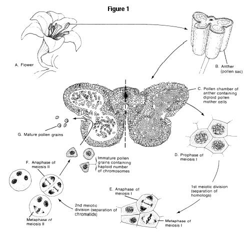
1. MEIOSIS IN FLOWERING PLANTS
You will study meiosis as it occurs in the development
of mature pollen grains of flowering plants. These pollen grains give rise
to male gametes (sperm nuclei), which fuse with an egg to produce a zygote.
As you examine the series of slides in the meiotic sequence, refer to figure
1 below for help in locating the stages.
a Meiosis I
Examine the illustration of the lily flower below,
figure 1, and locate the anthers, or pollen sacs, which contain numerous
microspore mother cells (or microsporocytes). These cells undergo meiosis
and produce microspores. Eventually these microspores develop into pollen
grains, which in turn produce sperm.
What is the significance of this process?______________________________________________________________________________________________________
______________________________________________________________________________________________________
During metaphase I, the homologous chromosomes move toward the spindle equator. The centromeres of each homologue come to lie on either side of the equator. One homologue becomes attached to spindle fibers extending from 1 pole, and the other homologue becomes attached to fibers extending from the opposite pole.
How does this differ from the alignment you observed in cells going through metaphase of mitosis?__________________________________________________________
______________________________________________________________________________________________________
During anaphase I, the homologous separate, 1 homologue
moving toward
1 pole, the other toward the opposite pole. Each homologue
is still in the duplicated form, consisting of 2 sister chromatids, attached
by their centromere. Examine slides of a lily anther showing separation
of homologous chromosomes (Fig. 1E).
What was the status of the sister chromatids during anaphase of mitosis?________________________________________________________________________________
______________________________________________________________________________________________________
Meiosis I is completed at the end of telophase 1. As cytokinesis begins, a cell plate forms in the midplane of the cell, and a nuclear envelope reforms around the chromosomes, resulting in a well-defined nucleus in each of the 2 daughter cells. Depending on the organism involved, an interphase may precede meiosis II, during which a cell wall forms across the entire, of the midplane. It is important to note here that DNA synthesis does not occur following telophase I (between meiosis I and meiosis II).
b. Meiosis II
At the beginning of the 2nd meiotic division (prophase
II), the sister chromatids are still attached by their centromeres. During
metaphase II, the duplicated chromosomes align on the spindle equator,
with the centromeres lining on the equator. (Metaphase II of meiosis looks
very similar to metaphase of mitosis.)
During anaphase II (Fig. 1F), the centromeres split,
and the chromatids that make up each chromosome now separate and migrate
to opposite poles. As in mitosis, the chromatids, when separated from each
other, are called daughter chromosomes. At the poles, during telophase
II, the daughter chromosomes become enclosed in a nuclear envelope. Cytokinesis
follows the division of the nucleus.
After cell wall formation is complete, the 4 haploid
microspores will separate. Subsequently, each will develop into a pollen
grain, inside which sperm cells will be formed.
2. MEIOSIS IN ANIMALS
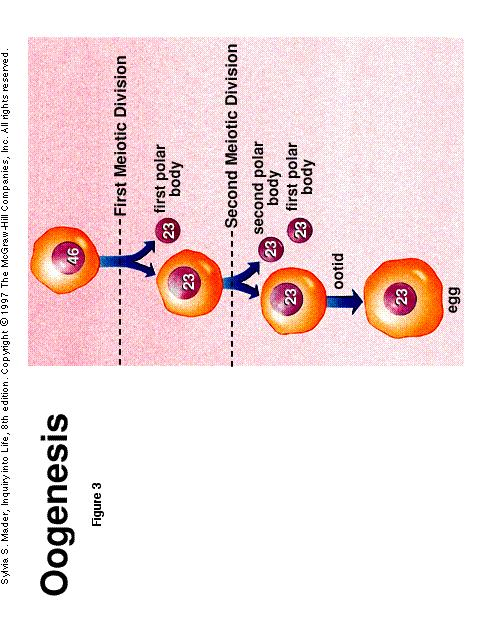
The formation of gametes is known as gametogenesis; the production of sperm is referred to as spermatogenesis; production of eggs or ova is referred to as oogenesis. In animals, gametes are produced in the gonadsovaries and testes. In fact, this is the only place where meiosis occurs in higher animals. In this part of the laboratory exercise, you will study the meiotic events in oogenesis as they occur in mammals. Refer to the attached figures 2 and 3 of oogenesis and spermatogenesis.
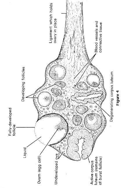
The cells in the ovary that give rise to female gametes are called primary oocytes; these cells are diploid. As in all vertebrate animals, mammalian primary oocytes are surrounded during their entire growth and maturation stages by follicle cells (or granulosa cells), which secrete substances which assist the growth of the oocyte. The oocyte and its mass of follicle cells are collectively known as a follicle. Examine slides containing mammalian ovaries. Refer to figure 4 attached. You will probably first see several follicles at different stages of development. The larger ones have a clear space, the antrum, within the mass of follicles cells, which under normal circumstances is filled with follicular fluid. The less mature follicles are not only smaller, but may even appear solid due to the fact that the antrum has not yet formed. Each human follicle contains an oocyte, which may not be seen, due to the fact that in sectioning the ovary, the oocyte did not lie in the plane that was cut. In dog follicles, there may be as many as 5 oocytes within a single follicle.
Prior to the onset of oogenesis, the chromosomes of each primary oocyte replicate to form sister chromatids, and then the primary oocytes begin to undergo prophase I of meiosis. During prophase I, the pairs of homologous chromosomes lie adjacent to each other (synapsis) and form tetrads. Crossing over usually occurs at this time, which results in an exchange of genetic components between the homologous. Locate primary oocytes in which synapsis is taking place.
During anaphase I, the members of the homologous pairs of chromosomes, each homologue still consisting of a pair of sister chromatids, move to opposite poles. Since the centromeres do not separate, the sister chromatids remain together.
During telophase I, an unequal division of the cytoplasm occurs. This results in a large cell called a secondary oocyte and a small cell called the first polar body. Only the secondary oocyte will go on to become a mature, haploid ovum; depending on the species, the polar body may or may not undergo meiosis II. In any case, the polar bodies are extremely small and do not function as gametes. Locate the first polar body on the slide.
How many chromosomes are found in the first polar body?_____________________
How many chromosomes are found in the secondary ooctye?___________________
Prophase II is essentially a repeat of prophase I, The homologous, however, have now been separated into the 2 secondary oocytes. In metaphase II, each single chromatid now lines up on the metaphase plate. This stage looks remarkably like metaphase in mitosis, since there is no pairing of the homologous. The second meiotic division produces both a second polar body and a large cell that differentiates into the ovum, or egg. (The first polar body produced in meiosis 1 may or may not got through a 2nd meiotic division.) Thus, when a diploid cell in the ovary undergoes complete meiosis, only 1 mature ovum is produced; the polar bodies are essentially nonfunctional. The unequal cytokinesis during oogenesis ensures that an unusually large supply of cytoplasm and stored food is allotted to the nonmotile ovum for use by the embryo that will develop from it. In fact, the ovum provides all of the cytoplasm and initial food supply for the embryo. The tiny, highly motile sperm cell contributes only its genetic material.
In mammals, oogenesis is characterized by periods
of meiotic arrest. In the human female, meiosis I begins in the embryo,
as early as the fourth month of life, but meiosis is not completed until
onset of puberty (at the earliest) or just before menopause (at the latest).
The first arrest occurs during prophase I; this arrest can last 40 years
in some human ova. At the onset of puberty, circulating hormones stimulate
growth of 1 or 2 of these dormant follicles (and their primary oocytes)
each month. The oocyte will enlarge and the number of follicular cells
increases. Meiosis in the primary oocyte resumes, and meiosis I is completed,
resulting in one secondary oocyte and the first polar body. But meiosis
is arrested again, usually in metaphase II. When The secondary oocyte is
released from the ovary, along with many of the follicular cells (ovulation,
Fig. 10); meiosis II is completed only after the cell is fertilized by
a sperm. Completion of meiosis II produces another polar body.
After ovulation, the follicle collapses and becomes
transformed into a small endocrine organ known as the corpus luteum (Greek,
"yellow body"). The corpus luteum produces hormones, especially progesterone,
that prepare the uterus for the potential arrival of a fertilized egg.
3. MEIOSIS COMPUTER LAB
For this part of the lab you will be using the program Meiosis by the EME Cooperation. It can be found in the computer lab in the Gen.Bio. Human folder.
MEIOSIS COMPUTER LAB
1) This shows how, in the process of meiosis, the number of chromosomes is reduced by half in the formation of gametes. An understanding of how chromosomes duplicate and divide during mitotic cell division is essential before using the program. Study the diagrams of mitotic stages below. Italicized terms appear in the Glossary attached.
2. When the program has been loaded into the computer,
run the INTRODUCTION for background on the process of meiosis.
Activity I
Run the MEIOSIS SIMULATION in normal spermatogenesis and normal oogenesis. Then answer the following questions:
A. What is the relationship between a chromosome
and a chromatid ?
B. Why does crossing over occur only during synapsis
?
C. In the model, the chromosomes from the male parent
are color coded blue, those from the female parent white. Since there
are 6 chromosomes,3 will be blue and 3 will be white. Does
this mean in oogenesis that there will be 3 white chromosomes in the egg?________
NOTE: In the IBM version the male chromosomes are green and the females
are red. Will there be 3 red chromosomes in the egg? ______
Why or why not?
D. What distinguishes the second stage of meiosis
from mitosis?
E. Describe how oogenesis differs from spermatogenesis.
Activity II
When male and female games unite a zygote is formed. The zygote has a diploid (2N) number of chromosomes because it results from 2 haploid (N) cells uniting. The fertilized egg becomes the new individual.
A. What would happen to the chromosome number in
gametes if they were formed by the mitotic process instead of the meiotic
process?
B. How are the chromosomes in the zygote related
to the parents of the male and female whose gametes united to form it?
Chromosomes bear the traits of the organism encoded in
the form of DNA nucleotide sequences and they are transmitted in
the gametes from one generation to the next. Instructions for specific
traits are located at sites on the chromosomes called genes. Strands
of DNA that make up chromosomes may consist of hundreds or thousands of
genes. Homologous chromosomes have genes for the same trait
located at the same site.
C. What might happen to a gene during crossing over?
D. What would happen to the traits if homologous
chromosomes did not come together as tetrads?
Activity III
Run the simulation in nondisjunction for spermatogenesis and oogenesis.
A. When nondisjunction occurs during spermatogenesis how many spermatids will be: normal________ , exhibit trysomy (one chromosome in triplicate) _______ , exhibit monosomy (single instead of a pair of chromosomes)________?
B. If nondisjunction occurs during Anaphase II of
oogenesis what are the possible effects on the ootids N number?
Activity IV
Draw and label the stages of oogenesis and spermatogenesis
below.
GLOSSARY
centromeres: the point at which identical or sister chromatids are joined together.
chromatid: one strand of a chromosome after it has been replicated.
chromatin: collectively, the decondensed interphase chromosomes.
chromosomes: strands of DNA that are the genetic material of the cell.
crossing over: the process, occurring during synapsis, in which homologous chromosomes exchange segments of chromosome.
cytokinesis: division of the cell's cytoplasm.
diploid: two complete sots of chromosomes, (2N)
gamete: male or female reproduction cell.
gene: a portion of a DNA molecule that codes hr a product such as a particular protein.
haploid: half the diploid number (N), or one complete set of chromosomes.
homologous chromosomes: those containing genes for the same traits.
meiosis: duplication and division of chromosomes resulting in gametes with half the normal number of chromosomes.
mitosis: process of nuclear division that results in two new genetically identical nuclei.
monosomy: condition in which a cell contains only one member of a chromosome pair instead of the usual two.
nondisjunction: the failure of a pair of chromosomes to separate during meiosis.
oogenesis: meiosis resulting in ootids.
ootid: (N) cell produced during oogenesis that will mature into an egg.
polar body: (N) cell produced during oogenesis that does not mature.
primary oocyte: (2N) cell in prophase I of oogenesis that will result in a secondary oocyte and first polar body.
secondary oocyte: (N) cell in prophase II of meiosis that will result in an egg and second polar body.
somatic: a body cell with 2N number of chromosomes.
spermatid: (N) cell produced during spermatogenesis that will mature into a sperm
spermatogenesis: meiosis resulting in spermatids.
synapsis: stage in prophase I of meiosis where homologous chromosomes come into contact.
tetrad: two homologous chromosome four (chromatids) in synapsis.
trisomy: condition in which a cell contains three copies of a single chromosome instead of the usual two.
zygote: a fertilized egg